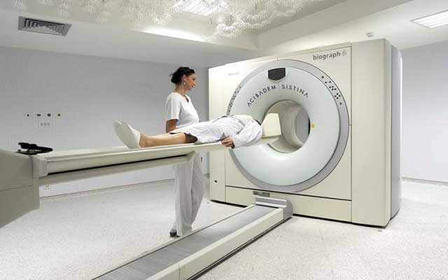
Positron emission tomography (PET) and computed tomography (CT) are combined into one machine in this advanced nuclear imaging technique. A lot of information about both the structure and function of the cells and tissues in the body during a single imaging session is revealed by a PET/CT scan. The patient is first injected with a glucose solution containing a very small amount of radioactive material during a PET/CT scan. The particular organs or tissues being examined absorb the substance. On a table, the patient rests and it slides into a large tunnel-shaped scanner. Then the damaged or cancerous cells where the glucose is taken up can be seen by the PET/CT scanner. The rate at which the glucose is used by a tumour can also be seen by the scanner. The tumour grade can be determined by this, which helps in cancer treatment. The patient does not feel any pain in this procedure, which varies in length depending on the body part that is being evaluated. A PET/CT scan provides a more detailed picture of cancerous tissues than either test does alone as it combines information about the body’s anatomy and metabolic function. In a single scan, the images are captured resulting in a high level of accuracy. Prior to ordering a PET/CT scan, an oncologist will often perform a CT scan and/or a bone scan.
How is the procedure performed?
On an examination table, you will be positioned and a nurse or technologist will insert an intravenous (IV) catheter into a vein in your hand or arm. The dose of radio tracer is then injected intravenously, swallowed or inhaled as a gas depending on the type of nuclear medicine exam you are undergoing. The time taken by the radio tracer to travel through your body and to be absorbed by the organ or tissue being studied will be approximately 60 minutes. To help the radiologist interpret the study, you may be asked to drink some contrast material that will localize in the intestine. The imaging will begin when you will be moved into the PET/CT scanner. During imaging, you need to remain silent. The PET scan will be done after the CT scan. Sometimes, after the PET scan, a second CT scan with intravenous contrast may be done. Nearly 30 minutes will be the total scanning time. You may be asked to wait after the examination is done until the technologist checks the images in case additional images are needed. In some cases, for clarification or better visualization of certain areas or structures, more images are obtained. The cancer doctor can do better diagnosis if the images are clear. You should not be concerned if additional images are needed because that does not necessarily mean that something abnormal was found or there was a problem with the exam. For the procedure, if an intravenous line was inserted in your body, it will usually be removed unless an additional procedure is needed the same day that requires an intravenous line.
Uses
There are various uses of whole body PET/CT scan. The following are some of them:
- Metastatic work up that is to ascertain the tumor load
- Response evaluation in cases where chemotherapy is used as first line of treatment
- To determine recurrence after completion of therapy
Contraindication
- For brain tumours
- Thyroid cancer
Relatively contraindication
- Uncontrolled diabetes
- Immediately after completion of therapy

